In the late 1970s and early 1980s, arthroscopic surgery became popular, especially in the sports world, as fibre optic technology enabled surgeons to see inside the body using a small telescope, called an "arthroscope", which projects an image to a television monitor.
Thanks to on-going improvements, the benefits of arthroscopic surgery for knee and shoulder conditions have been experienced by patients all over the world. By adopting techniques and instruments similar to those used in knee and shoulder procedures, arthroscopic hip surgery has become a more widely-used treatment option for those who suffer from hip pain.
Arthroscopic procedures may be used for a variety of hip conditions, primarily the treatment of hip impingement, labral tears, articular cartilage injuries, the removal of loose bodies in the joint and snapping hip syndrome. Other less frequent conditions treated through hip arthroscopy include tendon or ligament injuries, hip instability, and an inflamed or damaged synovium.
Through an incision the width of a straw tip, a surgeon is able to insert a scope, which allows him or her to inspect the joint and locate the source of pain. The surgeon will then make one or more small incisions to accommodate the instruments used to treat the hip. These instruments can shave, trim, cut, stitch, or smooth the damaged areas.
Hip arthroscopy is less invasive than traditional open hip surgery, is often performed as a day-case and may require a shorter stay in hospital compared to traditional open hip surgery.
what to expect
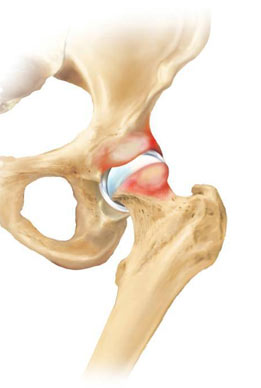
tophip impingement
With hip impingement treatment, the surgeon will reshape the junction between the head and neck of the femur using small mechanical resection devices called burrs. Performing this step as well as trimming any excessive portion of the acetabulum will give the joint more clearance, thus relieving the impingement. At various times during the surgery and immediately following it, the surgeon will test and monitor the range of motion of the hip.
labral tears
In this procedure, the surgeon will smooth the edges of the torn or frayed labrum using arthroscopic shaver blades or radiofrequency (RF) energy. Specially designed RF probes include flexible heads that allow the surgeon to manoeuvre through difficult curves in the hip joint, remove torn tissue and smooth the damaged areas. In some cases, the labrum may be repaired. For this procedure, anchors will be attached to the bone and sutures will be passed through the tissue. The anchors are used to hold the suture in place.
articular cartilage injuries
To treat articular cartilage injuries, the surgeon will use an arthroscopic shaver blade to remove the damaged tissue, leaving a smooth stable surface. Certain types of injuries may require treatment with microfracture. In this procedure, the surgeon will create a number of small holes in the exposed bone of the joint to induce bleeding and clotting, which also leads to new tissue growth. Studies indicate that in time, this new growth becomes firm tissue that is smooth and durable.
loose bodies
When removing loose bodies, the surgeon will use an arthroscope to inspect the joint to confirm the number of loose bodies and their location. The surgeon will then remove the loose bodies using specially designed hand instruments called graspers.
snapping hip syndrome
To treat snapping hip syndrome (extra-articular lesions), the surgeon must release the snapping iliopsoas tendon, or less commonly, the tensor fascia lata. During the procedure, the surgeon will rotate the hip and make two portals; one for the arthroscope and one for a specially designed radiofrequency (RF) probe. The RF probe has a flexible head, allowing the surgeon to release the tendon. This method is safer than open surgery, and studies have shown that there are minimal complications following the procedure4
pre-surgery
topPreparation for surgery begins weeks and sometimes months before the surgery date and may include:
initial surgical consultation
Preoperative X-rays, a complete medical history, a complete surgical history, and a complete list of all medications (i.e. prescription, over- the- counter, vitamin supplements) and allergies will be reviewed.
complete physical examination
The surgeon will perform a physical examination and work with a patients doctor if required to manage other medical conditions.
physiotherapy
Instruction in an exercise program to begin prior to the surgery, as well as an overview of the rehabilitation process after surgery, will better prepare you for post-operative care.
personal preparation
Loose fitting clothing is recommended. Patients should bring a list of all medications, dosages, and any known allergies.
evening before surgery
The surgeon may recommend not eating or drinking after midnight. The surgeon or anaesthetist may also recommend taking routine prescription medications with a sip of water.
On the day of surgery, a patient can expect:
- To be admitted to hospital.
- For their vital signs, such as blood pressure and temperature, to be measured.
- To be given a clean hospital gown.
- To remove all jewellery, dentures, contact lenses, and nail polish.
- An IV to be started to give fluids and medication during and after the surgery.
- The hip to be scrubbed and shaved in preparation for the surgery.
- An anaesthetist to discuss the anaesthetic that will be used.
- The surgeon to confirm and initial the correct surgical site.
the surgery
topThe techniques used during a hip arthroscopy will vary depending on the type of hip problem being treated.
treatment of hip impingement
With hip impingement treatment, the surgeon will reshape the junction between the head and neck of the femur using small mechanical resection devices called burrs. Performing this step as well as trimming any excessive portion of the acetabulum will give the joint more clearance, thus relieving the impingement. At various times during the surgery and immediately following it, the surgeon will test and monitor the range of motion of the hip.
treatment of labral tears
In this procedure, the surgeon will smooth the edges of the torn or frayed labrum using arthroscopic shaver blades or radiofrequency (RF) energy. Specially designed RF probes include flexible heads that allow the surgeon to manoeuvre through difficult curves in the hip joint, remove torn tissue and smooth the damaged areas. In some cases, the labrum may be repaired. For this procedure, anchors will be attached to the bone and sutures will be passed through the tissue. The anchors are used to hold the suture in place.
treatment of articular cartilage injuries
To treat articular cartilage injuries, the surgeon will use an arthroscopic shaver blade to remove the damaged tissue, leaving a smooth stable surface. Certain types of injuries may require treatment with microfracture. In this procedure, the surgeon will create a number of small holes in the exposed bone of the joint to induce bleeding and clotting, which also leads to new tissue growth. Studies indicate that in time, this new growth becomes firm tissue that is smooth and durable.
loose body removal
When removing loose bodies, the surgeon will first use the visibility provided by the arthroscope to inspect the joint. This inspection will help confirm the number of loose bodies and their location. The surgeon will then retrieve and remove the loose bodies using specially designed hand instruments called graspers.
treatment of snapping hip
To treat snapping hip syndrome (extra-articular lesions), the surgeon must release the snapping iliopsoas tendon, or less commonly, the tensor fascia lata. During the procedure, the surgeon will rotate the hip and make two portals; one for the arthroscope and one for a specially designed radiofrequency (RF) probe. The RF probe has a flexible head, allowing the surgeon to release the tendon. This method is safer than open surgery, and studies have shown that there are minimal complications following the procedure4.
recovery
topAfter surgery, patients are transported to a recovery room for close observation of vital signs and circulation.
The condition being treated will determine whether or not a patient will be required to use crutches when leaving the hospital and the length of time crutches may be needed. The doctor will provide specific information regarding a patients own specific post-operative care.
watch/listen